
Reflections of an image
Digital holography is a phase contrast microscopy technique that uses the interference of light waves to create an image. You can think of it in terms of waves on a beach: if there is a rock off-shore, refracting the waves around it, you could reconstruct the shape of that rock by measuring the pattern of waves hitting the sand.
With digital holographic microscopy, the waves are light beams and the rock is often a cell held suspended in a solution. By measuring the phase shift of light passing through the cell, a 3D holographic image of that cell can be reconstructed using a digital algorithm. That image can then be used to analyze a wide range of morphological parameters, such as cell volume and shape. Other important parameters can also be computed, including viability, viral load, cell cycle and apoptotic state. When scaled up, you can use this method to obtain accurate cell counts and viability data for an entire biological sample, non-invasively and in-real time.
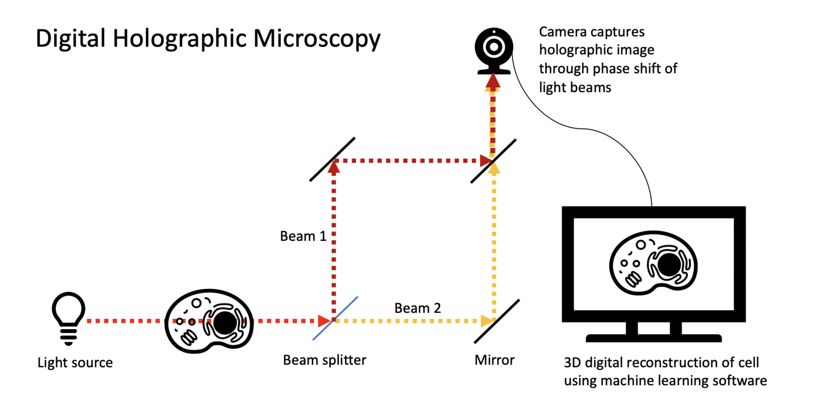
Image: Digital holographic microscopy works by using the phase shift patterns of light passing through an object to reconstruct a 3D image using AI.
Digital holographic microscopy is not common but there are some other companies using their own version of the imaging technique. However, these other companies usually use lasers as their coherent light source. This is adequate for material sciences, where the lasers are shone at inert objects, but presents problematic side-effects when applied to living cells. Ovizio’s technology differs from these laser-based devices in that they instead use partially coherent light, based on LEDs. This eliminates the complications, like phototoxicity, caused by more intense laser lights.
A solution for cells in suspension
Ovizio’s patented devices can be used in a range of applications, including hematology, material sciences and even the food industry. However, the Brussels-based team has developed the technology to address a specific challenge: the monitoring of cells in suspension. Currently, they are the only company in the world providing online monitoring solutions for suspension cell culture bioreactors.
Bioreactors are cell production factories used for many important biopharmaceutical processes, including vaccine manufacturing and CAR T-cell therapy. Ovizio’s digital holographic microscopes present a real game-changing alternative to the imaging methods traditionally used to check cell viability and proliferation in these therapeutic areas, such as phase contrast microscopy and Trypan blue stains.
The advantages of Ovizio’s technology are significant. Current monitoring practices typically involve off-line sampling: to measure cell viability, you need to extract cells from your sample via a syringe, stain those cells and put them through an off-line cell counter. This method immediately presents several major issues: you risk contamination by interfering with the cell culture, a huge concern if you’re working with cell therapy. Sterility cannot be compromised under any circumstances as the cells will ultimately be injected into a patient. If you are working with a very small initial sample, you are also losing cells, which is not ideal. Finally, the process is time and labor intensive and does not provide you with real-time data on how the cells in your sample are faring.
Ovizio’s technology provides a solution to all of the aforementioned issues. Digital holographic microscopy does not need cells to be stained to count them or measure their viability, so the procedure is label-free and non-invasive. The device works using a closed-loop system, meaning no sample is lost and there is no increased risk of contamination and compromising sterility. Finally, Ovizio engineers have automated the entire system, meaning you can obtain real-time information on your samples with a minimal time and energy investment.
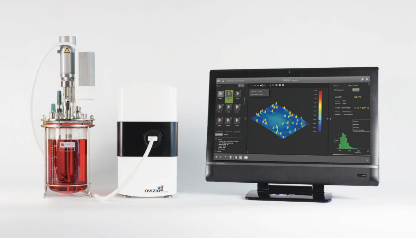
Image: One of Ovizio’s microscopes with a display showing a cell viability count.
An intelligent approach
At the core of Ovizio’s technology you’ll find an intelligent piece of software: a program based on machine learning that allows for the automation of the bioreactor monitoring process. This AI driven software can be taught to classify the holographic images obtained by the Ovizio microscopes into different categories, such as cell types, based on deep learning algorithms. The adaptive capabilities of the program also mean that Ovizio’s technology can be used for a wide variety of different applications and can be adjusted to different tasks simply by re-educating the computer. It also eliminates user bias caused by the slight (or not-so-slight) variations in results caused by the differences between individual technicians.
Ovizio have endeavored to make their microscopes as user friendly as possible, bringing their technology in line with the needs of a 21st century lab. The data extracted by their microscopes is remotely accessible, in real-time; much better than ordinary off-line bioreactor measurements, which typically require plenty of downtime and weekend work. By combining data acquisition and analysis with their machine learning software, Ovizio’s technology can save researchers both time and effort. This is especially important for a therapeutic area like CAR-T, where the manual labor costs for technicians drives up the price for what is already an exorbitantly priced therapy.
To achieve the full potential of new scientific solutions, like cell therapy, we often require new technological advances. Though phase contrast microscopy is a perfectly sound imaging method for obtaining qualitative data, the quantitative information provided by digital holography is more in-line with the needs of emerging therapeutic areas like CAR-T. In a fast-paced world, automation and refinement of data-gathering processes is key to pushing the limits of our medical capabilities.