You might also be interested in “Ending Alzheimer before it starts“
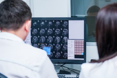
One out of six people in the world suffers from a neurological disorder, and 6.8 million people die from them every year. Chances are pretty high that someone in your family faces this problem. Many of us have a parent or a grandparent that suffers from dementia, which is often caused by Alzheimer’s disease (AD). We often use brain scans to measure the progression of neurological diseases. Every year, about 80 million MRI scans are taken, of which about 50% are used to get an image of the central nervous system (CNS). But do we process the information in those brain scans effectively?
Eye-balling vs measuring
To measure is to know. If you can not measure it, you can not improve it” – Lord William Thompson Kelvin
Let’s go to the history of measuring, where we clearly see that quantification has a huge impact on medicine. Blood glucose was initially measured based on the taste of urine. Luckily, we now have standardized glucose measuring devices that allow continuous monitoring of blood glucose levels, resulting in improved outcomes for diabetes patients. When it comes to brain scans, measuring is not yet widely implemented. A scan of a multiple sclerosis (MS) patient could be described, for example, as “having many lesions and moderate global atrophy.” This is a rather vague description and is not quantitative at all. We are still visually evaluating these images. There is an urgency to quantitate this. The measurement should be reproducible, accurate, sensitive, well-validated, objective and, preferably, cost efficient. In the ideal situation, a brain scan is analyzed by a computer, and you get a report with the data.
What’s your volume of brain loss?
A healthy subject has an estimated brain loss of 0.2% yearly where an AD patient has a loss of 1.5% yearly. Based on visual inspection, you cannot tell the difference in brain volume loss between 2 scans a year apart. Efforts have been made to quantitate changes with manual delineations, but this is very time-consuming, and there is a big inter-observer variability. Currently, there’s a shift toward 3D data, making manual delineations even more difficult.
At Icometrix, based in Leuven, they developed software for quantitative image analysis in areas such as brain atrophy. This product received CE approval and FDA clearance. Their software is sensitive, accurate and validated. For whole brain atrophy, the measurement error is 0.1%, which is small enough to determine the difference in brain loss between a healthy patient (0.2%) and an AD patient (1.5%).
Giving neurologists a hand
Let’s have a look at a few case studies. From 2011 onwards, an MS patient was on a disease modifying treatment. The patient had no new relapses since then. Nevertheless, the neurologist had the feeling that the patient was getting worse. But the neurologist was not able to derive clinical evidence from the clinical report. Then he looked into a quantitative MRI analysis report from Icometrix, which clearly confirmed what the neurologist was thinking: The brain volume was decreasing over time.
Icometrix’s software was also used to evaluate the long-term effect of natalizumab, a drug to reduce lesions in MS patients. They analyzed 1000 scans from 152 MS patients, including follow-up scans that were taken every year for five years. From these data, they could confirm that the lesions remained reduced for a longer period of time thanks to natalizumab.
We should stop eye-balling MRI images and start performing thorough, quantitative analyses of these scans. This could massively improve the treatment of neurological diseases.
This article was drafted based on the keynote presentation of Annemie Ribbens, Research Manager at Icometrix, at the FlandersBio–Janssen Pharmaceuticals Partner Day on March 9, 2017, at the University of Antwerp.